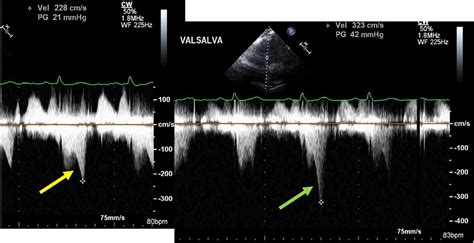
lv intracavitary gradient | intracavitary gradient is present lv intracavitary gradient Left ventricular cavity obliteration (LVCO), defined as obliteration of the apex in systole on angiography, was first described 1 in 1965 and proposed as the cause of the intraventricular pressure gradient accompanying hypertrophic cardiomyopathy. Genesect Pokémon Serebii.net Pokédex providing all details on moves, stats, abilities, evolution data and locations for Pokémon Sword & Shield
0 · normal Lv outflow gradient
1 · midcavitary gradient on echocardiogram
2 · mid cavitary gradient echo
3 · intracavitary gradient tte
4 · intracavitary gradient on echocardiogram
5 · intracavitary gradient is present
6 · intracavitary gradient echo
7 · Lv mid cavitary gradient
Enjoy German build quality from £5k with a 2004 Geist LV – it might take a while to find one on the used caravans for sale pages, but they're built to last. By Andy Jenkinson. Published: April 19, 2017. Geist caravans were solid, with very few suffering from damp issues.
Left ventricular cavity obliteration (LVCO), defined as obliteration of the apex in systole on angiography, was first described 1 in 1965 and proposed as the cause of the intraventricular pressure gradient accompanying hypertrophic cardiomyopathy.Hemodynamically, LVOTO has been defined as a peak instantaneous .We provide an overview of the pathophysiology of intraventricular . LVOTO is associated with impaired stroke volume and an increased risk of HF and poorer survival. 6,7 The presence of a peak LVOT .
The LVOT gradient is then estimated by the formula estimated LVSP – systolic BP, which reveals the correct answer choice of 130 mm Hg (275 – 145 mm Hg). Accurate measurement of the LVOT gradient is critical in the .The spectral profile of patients with AS, HOCM, and LVCO is differentiated by the peak/mean .
Hemodynamically, LVOTO has been defined as a peak instantaneous gradient at LV outflow of at least 30 mmHg, either at rest or on provocation. While traditionally defined in patients with hypertrophic .
We provide an overview of the pathophysiology of intraventricular obstruction induced by .Intracavitary gradients with cavity obliteration have been demonstrated during dobutamine . The spectral profile of patients with AS, HOCM, and LVCO is differentiated by the peak/mean gradient ratios of 2 or less, 2–3, and 3 or greater, respectively, in >90% of patients. Most patients. The primary hemodynamic effect on the left ventricle is one of increased afterload, resulting in increased intracavitary pressure and wall stress. In accordance with La Place’s law, the ventricle hypertrophies in an attempt to .
Left ventricular cavity obliteration (LVCO), defined as obliteration of the apex in systole on angiography, was first described 1 in 1965 and proposed as the cause of the intraventricular pressure gradient accompanying hypertrophic cardiomyopathy.Clinically significant LVOTO is often defined on the basis of echocardiography that demonstrates a pressure gradient across the LV outflow tract of >30 mm Hg. LVOTO is associated with impaired stroke volume and an increased risk of HF and poorer survival. 6,7 The presence of a peak LVOT gradient of ≥30 mm Hg is considered to be indicative of obstruction, with resting or provoked gradients ≥50 mm Hg generally considered to be the threshold for septal reduction therapy (SRT) in those patients with . The LVOT gradient is then estimated by the formula estimated LVSP – systolic BP, which reveals the correct answer choice of 130 mm Hg (275 – 145 mm Hg). Accurate measurement of the LVOT gradient is critical in the diagnosis and management of HOCM.
The spectral profile of patients with AS, HOCM, and LVCO is differentiated by the peak/mean gradient ratios of 2 or less, 2-3, and 3 or greater, respectively, in >90% of patients. Most patients with LVCO without HOCM or severe LVH have an ICG < 36 mm Hg.
Hemodynamically, LVOTO has been defined as a peak instantaneous gradient at LV outflow of at least 30 mmHg, either at rest or on provocation. While traditionally defined in patients with hypertrophic cardiomyopathy, LVOTO is known to have several causes.We provide an overview of the pathophysiology of intraventricular obstruction induced by exercise, highlighting its determinants: preload, contractility, and gradient. We describe the main signs of dynamic obstruction on echocardiographs.Intracavitary gradients with cavity obliteration have been demonstrated during dobutamine stress echocardiography and have, paradoxically, been associated with favorable, rather than adverse, outcomes 7,8 . The spectral profile of patients with AS, HOCM, and LVCO is differentiated by the peak/mean gradient ratios of 2 or less, 2–3, and 3 or greater, respectively, in >90% of patients. Most patients.
The primary hemodynamic effect on the left ventricle is one of increased afterload, resulting in increased intracavitary pressure and wall stress. In accordance with La Place’s law, the ventricle hypertrophies in an attempt to reduce wall stress.Left ventricular cavity obliteration (LVCO), defined as obliteration of the apex in systole on angiography, was first described 1 in 1965 and proposed as the cause of the intraventricular pressure gradient accompanying hypertrophic cardiomyopathy.Clinically significant LVOTO is often defined on the basis of echocardiography that demonstrates a pressure gradient across the LV outflow tract of >30 mm Hg. LVOTO is associated with impaired stroke volume and an increased risk of HF and poorer survival. 6,7 The presence of a peak LVOT gradient of ≥30 mm Hg is considered to be indicative of obstruction, with resting or provoked gradients ≥50 mm Hg generally considered to be the threshold for septal reduction therapy (SRT) in those patients with .
The LVOT gradient is then estimated by the formula estimated LVSP – systolic BP, which reveals the correct answer choice of 130 mm Hg (275 – 145 mm Hg). Accurate measurement of the LVOT gradient is critical in the diagnosis and management of HOCM.The spectral profile of patients with AS, HOCM, and LVCO is differentiated by the peak/mean gradient ratios of 2 or less, 2-3, and 3 or greater, respectively, in >90% of patients. Most patients with LVCO without HOCM or severe LVH have an ICG < 36 mm Hg.
Hemodynamically, LVOTO has been defined as a peak instantaneous gradient at LV outflow of at least 30 mmHg, either at rest or on provocation. While traditionally defined in patients with hypertrophic cardiomyopathy, LVOTO is known to have several causes.
We provide an overview of the pathophysiology of intraventricular obstruction induced by exercise, highlighting its determinants: preload, contractility, and gradient. We describe the main signs of dynamic obstruction on echocardiographs.
Intracavitary gradients with cavity obliteration have been demonstrated during dobutamine stress echocardiography and have, paradoxically, been associated with favorable, rather than adverse, outcomes 7,8 .
The spectral profile of patients with AS, HOCM, and LVCO is differentiated by the peak/mean gradient ratios of 2 or less, 2–3, and 3 or greater, respectively, in >90% of patients. Most patients.
burberry exploded check shirt

normal Lv outflow gradient
midcavitary gradient on echocardiogram

Creating a My LV account will enable you to: Save and manage your personal information. Create and share your personal wish list and bookmarks. Track your online orders. Receive our latest product and event updates. By clicking on My LV tab, you will be able to create your account and experience your personalised page on au.louisvuitton.com .
lv intracavitary gradient|intracavitary gradient is present


