lv noncompaction echo criteria | noncompaction cardiomyopathy criteria lv noncompaction echo criteria The objectives of this article are to review the imaging findings of left ventricular noncompaction (LVNC) at echocardiography, cardiac MRI, and MDCT; to discuss diagnostic . Kartē attēlota COVID-19 infekcijas izplatība Latvijas novados, pagastos, pilsētās un Rīgas apkaimēs.
0 · noncompaction cardiomyopathy guidelines
1 · noncompaction cardiomyopathy diagnosis
2 · noncompaction cardiomyopathy criteria
3 · left ventricular noncompaction syndrome
4 · left ventricular noncompaction radiology
5 · left ventricular noncompaction cardiomyopathy
6 · jenni criteria noncompaction
7 · jenni criteria lvnc
Lehigh Valley Zoo. 456 reviews. #1 of 6 things to do in Schnecksville. Zoos. Open now. 10:00 AM - 4:00 PM. Write a review. About. Lehigh Valley Zoo is home to approximately 300 animal ambassadors representing 104 species, 36 of which are classified as endangered, threatened, or species of concern. Meets animal welfare guidelines.
LVNC is characterized by the following features: An altered myocardial wall with prominent trabeculae and deep intertrabecular recesses, resulting in thickened myocardium . The objectives of this article are to review the imaging findings of left ventricular noncompaction (LVNC) at echocardiography, cardiac MRI, and MDCT; to discuss diagnostic .Diagnosis of noncompaction on echocardiographic criteria and at least 1 of the following: positive family history, associated neuromuscular disorder, regional wall motion abnormality, .
louis vuitton belt black monogram
A maximal endsystolic ratio of NC:C >2 has been established as one of the major criteria to diagnose LVNC by TTE and validated versus anatomical examination of the heart [1]. . CMR criteria to diagnose LVNC requires a higher non-compacted to compacted myocardium ratio than echocardiography, a ratio greater than 2.3 at end-diastole. CT can also provide an adequate definition of the increased .Left Ventricular Noncompaction Cardiomyopathy. Diagnostic Criteria. • Bilayered Bilayered myocardium myocardium (C+NC) (C+NC) • Ratio Ratio of of NC/C NC/C ≥ ≥ 2.0 2.0.2.1. Echocardiographic Criteria. Due to its low cost and widespread availability, 2D-echo is usually the first investigation in the evaluation of LV hyper-trabeculation. Presently, there are four 2D .
The three most commonly cited echocardiographic LVNC diagnostic criteria (Figure 1) measure the depths of intertrabecular recesses (Chin et al. criteria), the ratio of noncompacted to .
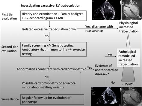
Left ventricular non-compaction (LVNC) is a rare congenital phenotype defined by the presence of prominent left ventricular trabeculae, deep intertrabecular recesses (continuous with the . Criteria for diagnosis by CMR: Petersen et al. (6) described the criteria for the diagnosis by CMR: the ratio of noncompacted myocardium to compacted myocardium must be greater than 2.3 during the diastole (sensitivity of 86% and specificity of 99%). LVNC is characterized by the following features: An altered myocardial wall with prominent trabeculae and deep intertrabecular recesses, resulting in thickened myocardium with two layers consisting of noncompacted myocardium and a thin compacted layer of myocardium (picture 1) [6-8].
The objectives of this article are to review the imaging findings of left ventricular noncompaction (LVNC) at echocardiography, cardiac MRI, and MDCT; to discuss diagnostic criteria for and the advantages and limitations of these imaging techniques; and to describe pitfalls that can lead to misinterpretation of findings of LVNC.Diagnosis of noncompaction on echocardiographic criteria and at least 1 of the following: positive family history, associated neuromuscular disorder, regional wall motion abnormality, noncompaction-related complications (arrhythmia, heart failure, or thromboembolism)A maximal endsystolic ratio of NC:C >2 has been established as one of the major criteria to diagnose LVNC by TTE and validated versus anatomical examination of the heart [1]. Compared to echocardiography, our CCT NC:C threshold of ≥1.8 NC:C is somewhat lower. CMR criteria to diagnose LVNC requires a higher non-compacted to compacted myocardium ratio than echocardiography, a ratio greater than 2.3 at end-diastole. CT can also provide an adequate definition of the increased trabeculations with less time and expense (but also less definition).
Left Ventricular Noncompaction Cardiomyopathy. Diagnostic Criteria. • Bilayered Bilayered myocardium myocardium (C+NC) (C+NC) • Ratio Ratio of of NC/C NC/C ≥ ≥ 2.0 2.0.
2.1. Echocardiographic Criteria. Due to its low cost and widespread availability, 2D-echo is usually the first investigation in the evaluation of LV hyper-trabeculation. Presently, there are four 2D-echo-based criteria that are commonly used, but none are considered as the gold standard (Table 1).The three most commonly cited echocardiographic LVNC diagnostic criteria (Figure 1) measure the depths of intertrabecular recesses (Chin et al. criteria), the ratio of noncompacted to compacted myocardium (Jenni et al. criteria), and the number of trabeculations (Stöllberger et al. criteria) [1,3–5].Left ventricular non-compaction (LVNC) is a rare congenital phenotype defined by the presence of prominent left ventricular trabeculae, deep intertrabecular recesses (continuous with the ventricular cavity), and a thin compacted layer.
Criteria for diagnosis by CMR: Petersen et al. (6) described the criteria for the diagnosis by CMR: the ratio of noncompacted myocardium to compacted myocardium must be greater than 2.3 during the diastole (sensitivity of 86% and specificity of 99%).
LVNC is characterized by the following features: An altered myocardial wall with prominent trabeculae and deep intertrabecular recesses, resulting in thickened myocardium with two layers consisting of noncompacted myocardium and a thin compacted layer of myocardium (picture 1) [6-8]. The objectives of this article are to review the imaging findings of left ventricular noncompaction (LVNC) at echocardiography, cardiac MRI, and MDCT; to discuss diagnostic criteria for and the advantages and limitations of these imaging techniques; and to describe pitfalls that can lead to misinterpretation of findings of LVNC.
Diagnosis of noncompaction on echocardiographic criteria and at least 1 of the following: positive family history, associated neuromuscular disorder, regional wall motion abnormality, noncompaction-related complications (arrhythmia, heart failure, or thromboembolism)A maximal endsystolic ratio of NC:C >2 has been established as one of the major criteria to diagnose LVNC by TTE and validated versus anatomical examination of the heart [1]. Compared to echocardiography, our CCT NC:C threshold of ≥1.8 NC:C is somewhat lower. CMR criteria to diagnose LVNC requires a higher non-compacted to compacted myocardium ratio than echocardiography, a ratio greater than 2.3 at end-diastole. CT can also provide an adequate definition of the increased trabeculations with less time and expense (but also less definition).Left Ventricular Noncompaction Cardiomyopathy. Diagnostic Criteria. • Bilayered Bilayered myocardium myocardium (C+NC) (C+NC) • Ratio Ratio of of NC/C NC/C ≥ ≥ 2.0 2.0.
2.1. Echocardiographic Criteria. Due to its low cost and widespread availability, 2D-echo is usually the first investigation in the evaluation of LV hyper-trabeculation. Presently, there are four 2D-echo-based criteria that are commonly used, but none are considered as the gold standard (Table 1).The three most commonly cited echocardiographic LVNC diagnostic criteria (Figure 1) measure the depths of intertrabecular recesses (Chin et al. criteria), the ratio of noncompacted to compacted myocardium (Jenni et al. criteria), and the number of trabeculations (Stöllberger et al. criteria) [1,3–5].
louis vuitton vintage monogram tote
noncompaction cardiomyopathy guidelines

louis vuitton monogram strass ring
noncompaction cardiomyopathy diagnosis
noncompaction cardiomyopathy criteria
CHILL.LV - AIM and FY Maps Only! Find the best CS:GO server by using our multiplayer servers list. Ranking and search for CS:GO servers.
lv noncompaction echo criteria|noncompaction cardiomyopathy criteria


